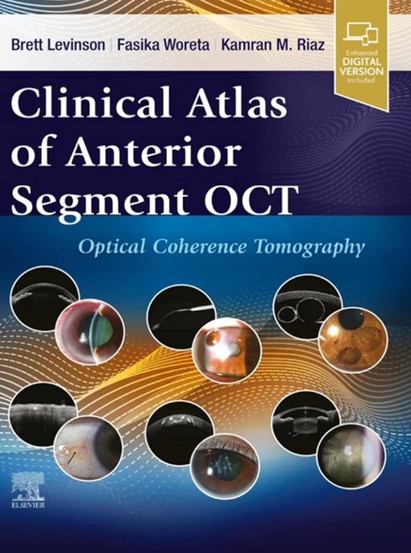
Clinical Atlas of Anterior Segment OCT: Optical Coherence Tomography
Brett Levinson, Fasika Woreta, Kamran Riaz, B0CVLNQ5RH, 0443120463, 0443120773, 9780443120466, 978-0443120466, 978-0-443-12046-6, 9780443120770, 978-0443120770
English | 2025 | PDF | 223 MB | 322 Pages
While eye care providers are thoroughly familiar with the use of optical coherence tomography (OCT) in the diagnosis and management of glaucoma and retinal diseases, many are not as familiar with its myriad uses for the diagnosis of corneal and anterior segment conditions. Anterior segment OCT (AS-OCT) can help to differentiate between various corneal pathologies, show the anatomy of the angles, obtain information about the lens-capsule complex, and guide contact lens fitting, among many other clinical uses. Clinical Atlas of Anterior Segment OCT expertly guides clinicians through all aspects of AS-OCT with hundreds of high-quality OCT images that highlight the utility of AS-OCT in diagnosing and managing a wide spectrum of anterior segment diseases.
- Covers the entire normal anatomy of the anterior segment and pathology of the conjunctiva, corneal epithelium, stroma and endothelium, lens, iris, anatomic angle, and clinical settings such as trauma, infection, inflammation, and contact lens fitting.
- Includes information on using AS-OCT in clinical and surgical settings (intraoperative AS-OCT).
- Provides rich visual guidance with over 500+ high-quality figures (anterior segment OCT imaging and clinical photos) comparing normal anatomy and a wide range of pathology, including both common and rare disorders and how to differentiate frequently confused conditions.
- Provides a well-rounded perspective of AS-OCT, including how to use, understand, and capture images.
- Links high-quality slit lamp images to the corresponding AS-OCT image with clear labels to show the pathology side by side.
- Features clinical pearls in each chapter to relate key AS-OCT and clinical findings to everyday practice.

